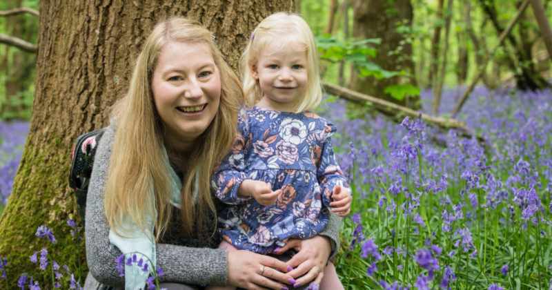
The prenatal scan that saved Emi’s life

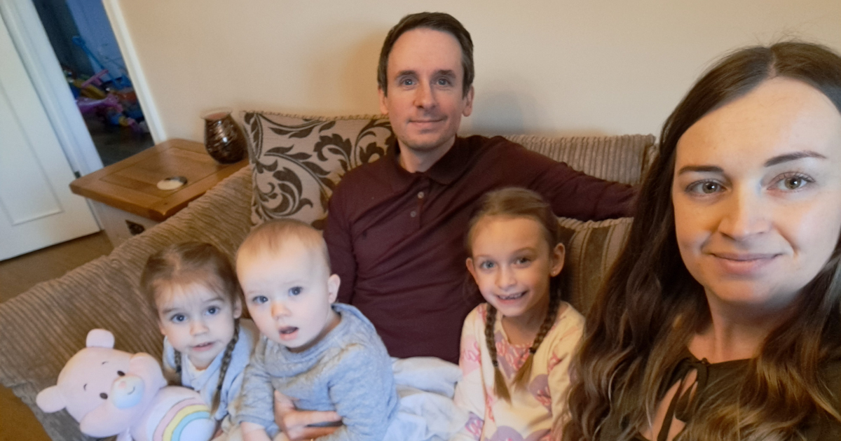
Louise King was full of excitement when she first saw her wriggly baby on the screen at her 13-week scan. But when the sonographer called in colleagues, she and her husband became concerned. They were told their baby had a life-threatening heart defect. Louise shares their story with MedTech Views on World Heart Day.
“When you hear there’s a problem with your baby’s heart, it rings alarm bells,” says Louise. “There was a lot of information to take in. The doctors drew a diagram and explained that the two main arteries of our daughter’s heart were swapped over.”
Their baby had a congenital condition called transposition of the great arteries (TGA). It meant that Emi’s pulmonary artery was where her aorta should be, and vice-versa. The pulmonary artery takes blood to your lungs to pick up oxygen and the aorta takes blood high in oxygen from your heart to your body. This meant that once Emi was born, she wouldn’t have enough oxygen moving around her body to survive.
“It was incredible that the scans picked up the defect even though Emi’s heart at that time was about the size of a grain of rice,” remembers Louise. “In addition to the ultrasound scans, we had a CVS [chorionic villus sampling] done to see if Emi’s condition was genetic or if she had any other genetic conditions, which she didn’t.” The CVS procedure involved a small sample of placental tissue being taken from Louise’s womb via a needle inserted into her abdomen, followed by analysis using other medical technologies.
There was also a small hole in the wall separating the two upper chambers (atria) of Emi’s heart, which was causing blood to flow abnormally between them. “A magnetic resonance imaging [MRI] scan allowed doctors to determine the size of the hole, so they knew that she would struggle to get enough oxygen as soon as she was born and they’d need to do a procedure straight away to allow for more oxygen flow,” Louise explains. “The balloon septostomy was done immediately via keyhole surgery.”
Using a medical technology called a balloon-tipped catheter (a special tube with a balloon at the end), surgeons were able to widen the hole to improve the blood flow and thus the oxygen levels in Emi’s heart. “Six days later, Emi underwent open heart surgery to swap over the arteries,” says Louise. “Throughout the whole journey, we had the support of the charity Tiny Tickers, which was a lifeline. We read through loads of babies’ stories, which were so helpful.”
“After the surgery, Emi spent five days on a ventilator because she had a partially collapsed lung, but she recovered quickly and was home when she was two and a half weeks old, after she’d gained back the weight that she’d lost,” notes Louise, who started volunteering for Tiny Tickers when Emi was 10 months old and working for them shortly after that. “Emi turned three last month. She has regular ultrasound scans that are getting less frequent. Now, they’re every 18 months and her heart is functioning normally.”
Louise wants to reassure other parents in this situation that they’re not alone. “There’s a lot of support. Seek out that support, read patient stories and don’t go through it alone.”